

內參抗體

- GAPDH Monoclonal Antibody
-
恒遠生物抗體平臺現已擁有4萬多種抗體(含單抗和多抗),包括人類常用抗體,模式生物類相關抗體、小分子抗體、二抗、標簽抗體及內參抗體等。并以每年4000種的速度在持續增長。
恒遠生物擁有專業的質控團隊,配備先進實驗儀器,現已成功地建立了ChIP平臺,因此我們的抗體不僅能提供Biotin、FITC、HRP等標記,大部分抗體都能進行ELISA、WB、IHC、IF、IP、ChIP等應用驗證。
我們致力于為所有用戶提供高親和力、高特異性的優質抗體。
-
貨號:HYK-200064
-
規格:
-
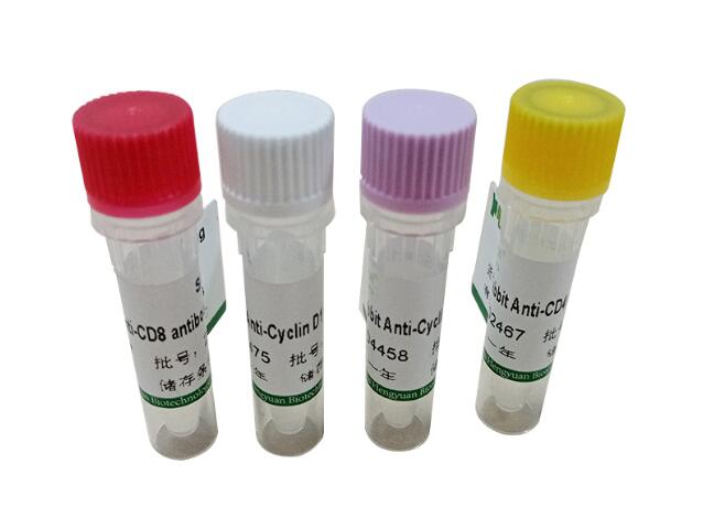
圖片:


產品詳情
-
產品名稱:Mouse anti-Homo sapiens (Human) GAPDH Monoclonal Antibody antibody
-
Uniprot No.:P04406
-
基因名:GAPDH
-
別名:GAPDH; G3PD; GAPD; MGC88685
-
宿主:Mouse
-
反應種屬:Human, Mouse, Rabbit
-
免疫原:Recombinant Human GAPDH protein (2-335AA)
-
免疫原種屬:Homo sapiens (Human)
-
標記方式:Non-conjugated
-
克隆類型:Monoclonal Antibody
-
抗體亞型:IgG1
-
純化方式:>95%, Protein G purified
-
克隆號:10B4E3
-
濃度:It differs from different batches. Please contact us to confirm it.
-
保存緩沖液:Preservative: 0.03% Proclin 300
Constituents: 50% Glycerol, 0.01M PBS, PH 7.4 -
產品提供形式:Liquid
-
應用范圍:ELISA, WB, IHC, IP, IF
-
推薦稀釋比:
Application Recommended Dilution WB 1:5000-1:80000 IHC 1:200-1:500 IF 1:50-1:100 IP 2μl-8μl -
Protocols:ELISA Protocol
Western Blotting (WB) Protocol
Immunohistochemistry (IHC) Protocol
Immunoprecipitation (IP) Protocol
Immunofluorescence (IF) Protocol
-
儲存條件:Upon receipt, store at -20°C or -80°C. Avoid repeated freeze.
-
貨期:Basically, we can dispatch the products out in 1-3 working days after receiving your orders. Delivery time maybe differs from different purchasing way or location, please kindly consult your local distributors for specific delivery time.
GAPDH Monoclonal Antibody
6*His Monoclonal Antibody
Myc tag Monoclonal Antibody
Sumo tag Monoclonal Antibody
MBP Monoclonal Antibody
GAPDH Monoclonal Antibody
在線詢價
- *
- *
- *
- *
- *
-
*
相關產品
Myc-Tag Monoclonal Antibody
恒遠生物抗體平臺現已擁有4萬多種抗體(含單抗和多抗),包括人類常用抗體,模式生物類相關抗體、小分子抗體、二抗、標簽抗體及內參抗體等。并以每年4000種的速度在持續增長。
查看詳細
HA-Tag Monoclonal Antibody
恒遠生物抗體平臺現已擁有4萬多種抗體(含單抗和多抗),包括人類常用抗體,模式生物類相關抗體、小分子抗體、二抗、標簽抗體及內參抗體等。并以每年4000種的速度在持續增長。
查看詳細
E-Tag Monoclonal Antibody
恒遠生物抗體平臺現已擁有4萬多種抗體(含單抗和多抗),包括人類常用抗體,模式生物類相關抗體、小分子抗體、二抗、標簽抗體及內參抗體等。并以每年4000種的速度在持續增長。
查看詳細